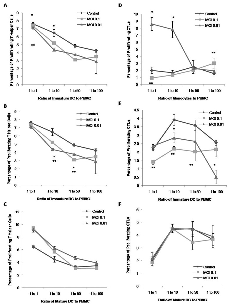
Figure 2. Immunostimulatory Capacity of Monocytes and Dendritic Cells Incubated with HCV.

Monocytes or DC from healthy human PBMC incubated with HCV were co-cultured with autologous PBMC containing CFSE for seven days at 1:1, 1:10, 1:50 or 1:100 ratio of effector cells to proliferators. Cells were then also stained for CD3, CD4 and CD8, and analyzed by flow cytometry. Figures are representative of three separate experiments (three separate donors). A) Percentage of proliferating helper T cells (CD3+ CD4+) after stimulation with monocytes. B) Percentage of proliferating helper T cells after stimulation with immature DC. C) Percentage of proliferating helper T cells with mature DC. D) Percentage of proliferating CTL (CD3+ CD8+) after stimulation with monocytes. E) Percentage of proliferating CTL after stimulation with immature DCs. F) Percentage of proliferating CTL after stimulation with mature DCs.
