
Figure 2.
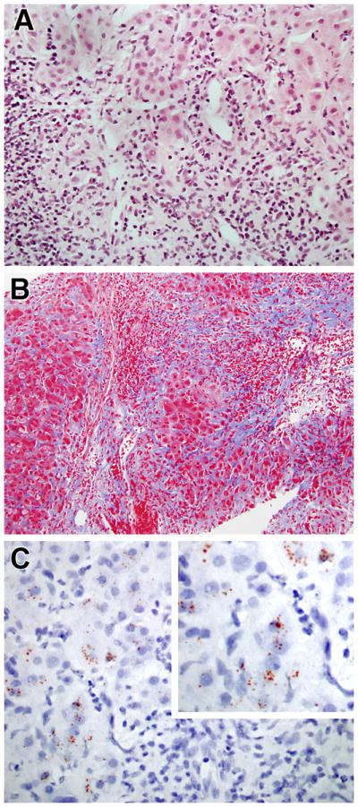
Progression of hepatic findings in CVID: A) Severe hepatitis with extensive interface hepatitis and focal bridging necrosis (H&E, 400x); B) Extensive fibrosis bridging portal areas (Masson trichrome, 200x); C) Rhodamine stain demonstrating red granules inside hepatocytes indicate of copper deposition (600x).