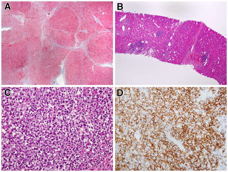
Figure 4.
Progressive liver disease and development of malignant lymphoma in a CVID patient with NRH: A) Masson trichrome staining showing incomplete cirrhosis with regenerative nodules partially and completely surrounded by fibrosis. (40x); B) H&E staining demonstrating lymphoid aggregates in portal areas and near central veins (100x); C) H&E staining showing late development of a large intra-hepatic lymphomatous mass (400x); D) Immunostain for CD20 demonstrating that the lymphomatous mass is largely composed of B cells (anti-L26, 400x)

