Fig. 5.
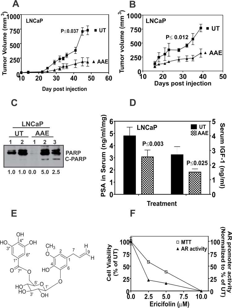
AAE inhibits in vivo growth of LNCaP in xenografts. (A) Growth of LNCaP tumors in male athymic nude mice with or without AAE gavage. Tumor volume was measured using hand-held microcalipers twice weekly as described in Materials and methods. Mice were gavaged daily, starting on the day of tumor cell injections for 50 days, with freshly prepared solution of AAE (100mg/kg). All tumor-bearing mice were euthanized 50 days after tumor cell injections. Symbols shown represent mean tumor volumes (vertical bars: SD, n = 8). (B) Growth of LNCaP tumors in athymic mice treated with AAE (50mg/kg by i.p. injection, 3×/week) or with saline injection (control). Experiment details are similar to that in Figure 5A. Mice were injected i.p. with AAE, three times a week at 50mg/kg starting from the day of tumor cell injection for 6 weeks. Significance of decreased tumor growth was tested by analysis of variance. The probability of observed difference in tumor volumes between AAE treated and control is due to chance is indicated (P ≤ 0.037 and P ≤ 0.012). (C) Expression of apoptotic biomarker, C-PARP in tumor tissues. Tumor tissues collected at necropsy were analyzed for C-PARP by extracting total proteins using a tissue homogenizer and lysing the tissue in NP-40 lysis buffer. Data shown for (D) serum IGF-1 and PSA levels in control and AAE-treated mice with LNCaP xenografts, IGF-1 and PSA levels in the sera of tumor-bearing mice were assayed using ELISA kits and expressed as ng/ml of serum. Data shown are mean ± SD (n = 3) for all treatments. (E) Identification of EF in AAE. EF was purified from AAE as described in Supplementary Data, available at Carcinogenesis Online. Approximately, 3.5mg of EF was purified from 1g of AAE; purity was confirmed using HPLC fractionation spectra followed by mass spectroscopy. Deduced chemical structure of eugenol-O-β-(6-galloylglucopyranoside) is shown. (F) EF purified from AAE inhibits cell proliferation and AR levels in LNCaP cells. LNCaP cells incubated with indicated concentrations of EF were assayed for cytotoxicity using MTT after 48 h. AR promoter reporter activity was assayed following 4 h treatment with EF, as described in Materials and methods. EF decreased viability of LNCaP cells (left ordinate) and AR transcriptional activity (right ordinate) (mean ± SEM, n = 5).

