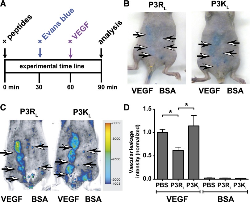
Figure 6.
TR47 decreases capillary vascular VEGF-induced permeability in the skin. VEGF-induced extravasation of Evans blue in the skin was determined in immune competent SKH1-E mice. (A) The experimental time line for the in vivo VEGF-induced vascular leakage model and administration of peptides. (B) Photographs for Evans blue extravasation in the skin of 2 representative mice that received the P3RL (left) or P3KL (right) peptide after subcutaneous injection with VEGF (3 left arrows) or with bovine serum albumin (BSA) control (2 right arrows). (C) Pseudo-colored heat map display of Evans blue extravasation quantified by the Odyssey near infrared imager at 700 nm. P3RL- (left) or P3KL- (right) treated mice were injected subcutaneously with VEGF (3 left arrows) or with BSA control (2 right arrows). (D) Quantification of in vivo VEGF-induced vascular leakage for mice treated with phosphate-buffered saline (PBS) control, P3RL (125 μg), or P3KL (125 μg) peptide. Data are also shown for BSA-treated control mice that received PBS, P3RL, or P3KL but no VEGF. (B-C) Representative experiments. (D) Data points represent the mean ± SEM (n ≥ 8). Asterisk denotes a statistically significant difference (P < .05).

