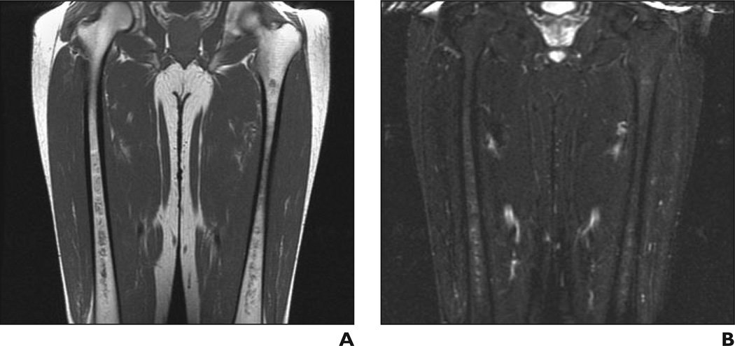
Fig. 5.
41-year-old man with Gaucher's disease.
A and B, Coronal T1-weighted (A) and STIR (B) MR images show minimal bone marrow infiltration with slightly low (A) and slightly high (B) signal intensity in both femurs with diaphyseal distribution. Bone marrow burden score is 3 (2 for intensity, 1 for distribution).

