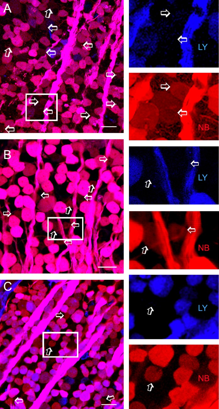
Figure 1.

ACs coupled to GC populations. Confocal micrographs from flat-mounted retinas from the wild-type mouse (A), conditional neuron-specific Cx45−/− mouse (B), and Cx36/Cx45−/− mouse (C) that were retrogradely double-labeled for LY (blue), and NB (red). A small population of displaced ACs (arrows and insets) is weakly positive for NB but negative for LY. The blue and red channels of each inset are separately depicted to the right. Coupled ACs show large to small soma size in wild-type mice, medium to small somas in Cx45−/− mice but mostly small somas in Cx36/45−/− mice. The number of coupled ACs is reduced more severely in Cx36/45−/− than in Cx45−/− mice. GCL, GC layer. Scale bar: 20 μm.
