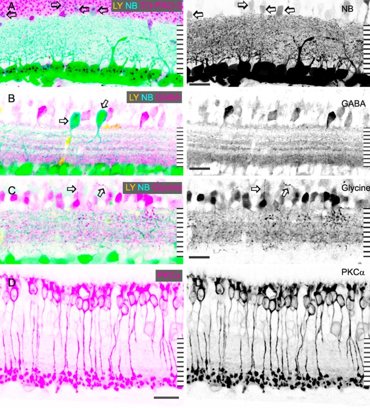
Figure 3.
The composition of the IPL. Confocal micrographs of retinal vertical sections that were retrograde-labeled by LY (yellow) and NB (blue), and further stained for TO-PRO-3 (A), GABA (B), glycine (C) or PKCα (D) in pink. For better clarity, some color channels are showed in black in the right panels. (A) is a stacked and overexposed image showing the distribution of LY/NB-positive GC dendrites in the IPL. Several GC-coupled ACs negative for LY but weakly positive for NB are visible in the INL (arrows, [A]), which are clearly distinguishable from displaced GCs (arrows, [B]). GC dendrites and glycine-IR are sparse in the inner 20% of the IPL (A, C), where PKCα-positive rod bipolar cell axon terminals (D) and the major GABA-IR band (B) are located. In the distal 80% of the IPL, GC dendrites, and glycine-IR are dense. Some AC somas in the GCL and INL are weakly PKCα-IR. Axon-like processes are emitted from some strongly glycine-IR somas (arrows, [C]). Scale bar: 20 μm.

