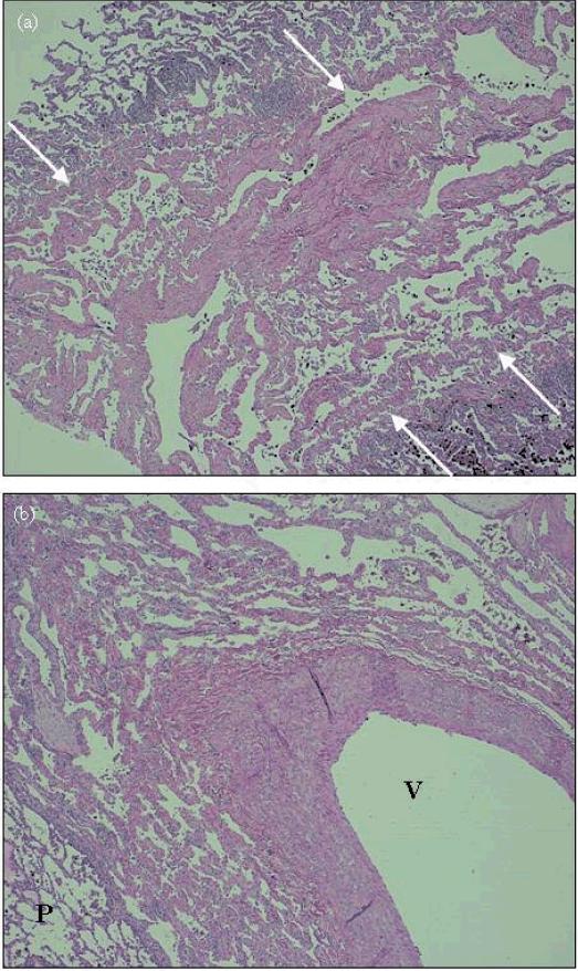
Figure 2.
H&E stain of native lung at 40x magnification. (a) right lung, peripheral upper lobe – septal lymphangiomatosis (between arrows) is seen in the center of the field, bordered on both sides by normal lung parenchyma. (b) left lung, central upper lobe – perivascular lymphangiomatosis, extensive lymphatic channels surround this arteriole (V indicates vessel, P indicated normal parenchyma).

