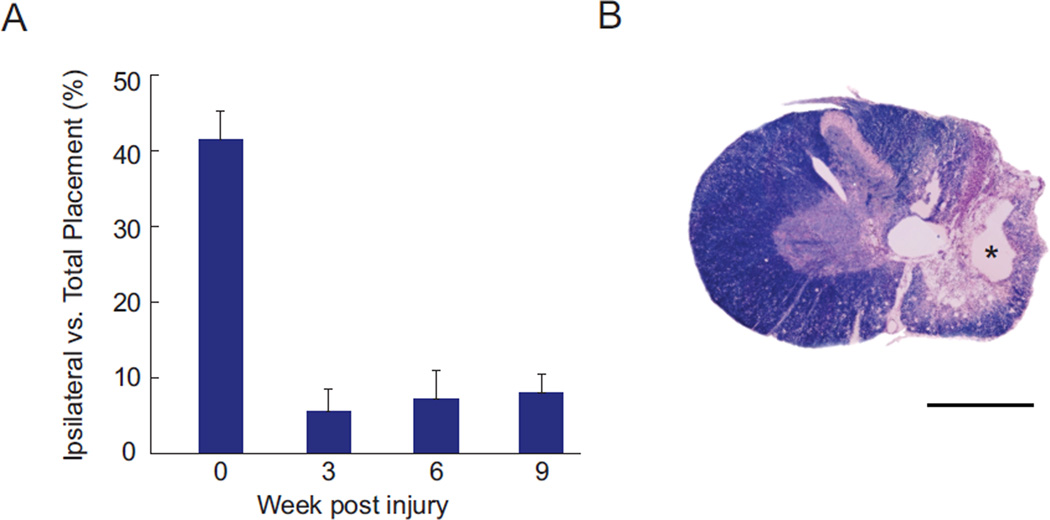
Figure 1.
Sustained forelimb deficits result from cervical contusion injuries. A: Forelimb asymmetry (cylinder) test demonstrates that animals use their ipsilateral to contusion forepaw for weight support less than 10% of the time after injury, compared to more than 40% of the time prior to injury (Mean ± SEM; N = 6 animals). B: Histology of a representative injury stained with cresyl violet (purple) and myelin stain (blue). Substantial cavitation is present at the injury epicenter (*), with wide-spread demyelination throughout the ipsilateral hemicord that persists for 20 weeks after injury. Scale bar in B is 1 mm.

