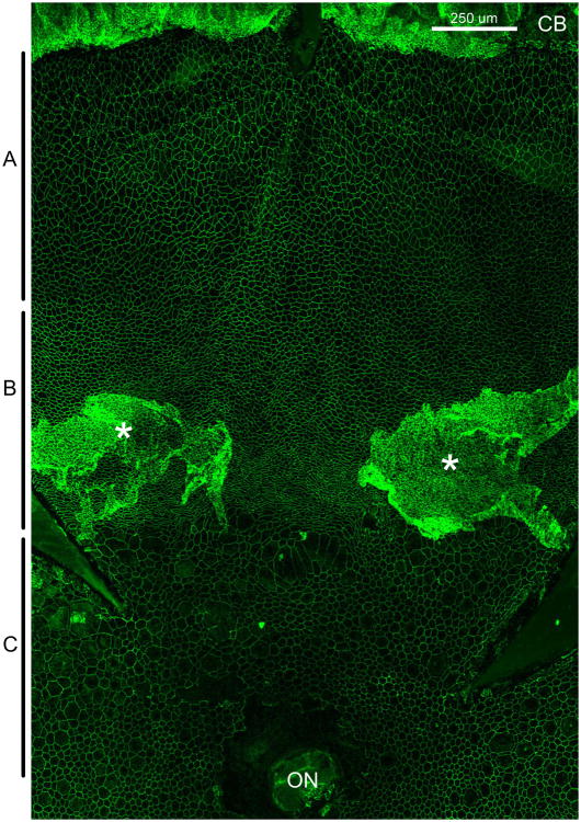
Fig.XX.1.
Superior flap of an RPE flatmount stained with ZO-1 to outline RPE cells from a 100 day old rd10 mouse. A indicates the peripheral region, B indicates the mid-periphery and C indicates the posterior region of the RPE sheet. CB – ciliary body, ON – optic nerve, * - areas of RPE ingrowth away from Bruch's membrane. Size bar is 250 μm.

