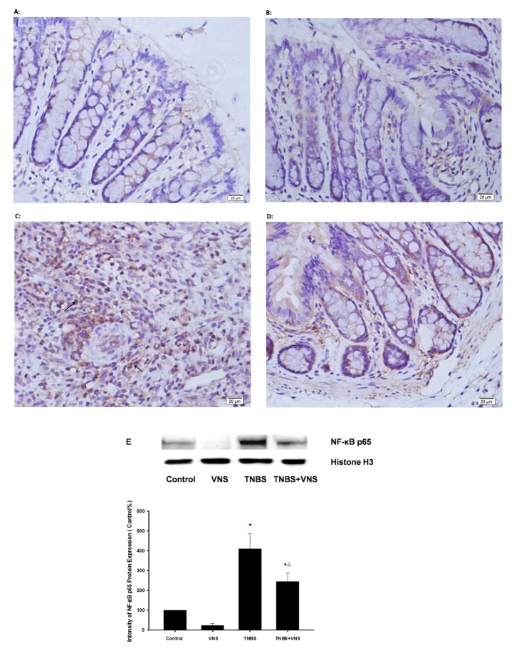
Figure 6. Chronic VNS inhibits the activation of NF-κB p65 on TNBS-induced colitis.
Photomicrographs (magnification ×400) are representative of immunohistochemically stained slides with NF-κB p65 anti-body in colon mucosa. (A) and (B) show normal colon mucosa, and colon mucosa with TNBS (C) shows markedly increased NF-κB p65 nuclear-positive cells (arrow). Colon mucosa from the TNBS model treated with VNS (D) shows much less translocation of NF-κB p65. Western blot was also performed with NF-κB p65 anti-body (E), and densitometric analysis was normalized to Histone H3. The results are expressed as the mean ± SEM (n = 3). The data shown are representative of three independent experiments. * P<0.05 versus the control group; Δ P<0.05 versus the TNBS group.

