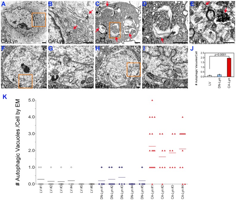
Figure 6. Expression of CA-Lyn in GBM cells results in larger tumors containing autophagic vacuoles.
Xenograft tumors expressing LV, CA-Lyn or DN-Lyn were generated as described in the legend for Figure 5 and after euthanasia on day 35 tumors were fixed in EM fixative, followed by transmission EM as described in the Materials and Methods. A-E, Three different representative cells from CA-Lyn tumors; panel B-higher magnification of the box in Panel A, and panel D-higher magnification of the box in panel C. F & G, Representative tumor cell from LV tumor; panel G-higher magnification of the box in panel F. H & I, Representative tumor cell from DN-Lyn tumor; panel I-higher magnification of the box in panel H. Scale bars in each panel denote 1 µm. Arrows denote examples of autophagic vacuoles. J, Quantitation of autophagic vacuoles/tumor cell shows a significant increase in the CA-Lyn tumors as compared to the LV tumors (p value <0.0001; t-test). K, Scattergram denoting the number of autophagic vacuoles/tumor cell.

