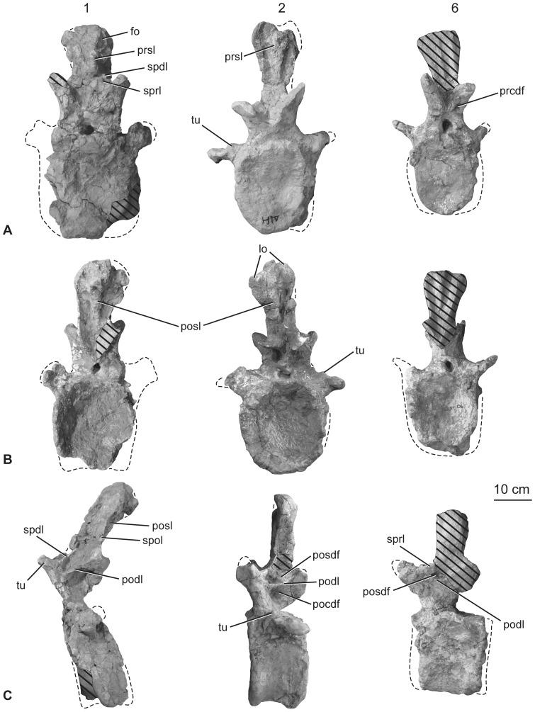
Figure 13. Holotypic anterior caudal vertebrae of H. allocotus (HBV-20001) from the Upper Cretaceous of Shanxi, China.
In (A) anterior; (B) posterior, and (C) left lateral views. First, second and sixth caudal vertebrae are depicted from left to right in each row. Lateral view of second caudal vertebra reversed. Abbreviations: di, diapophysis; fo, fossa; lo, lobe; tu, tubercle; pocdf; postzygapophyseal centrodiapophyseal fossa; podl, postzygodiapophyseal lamina; posdf, postzygapophyseal spinodiapophyseal fossa; posl, postspinal lamina; prcdf, prezygapophyseal centrodiapophyseal fossa; prsl, prespinal lamina; spol, spinopostzygapophyseal lamina; sprl, spinoprezygapophyseal lamina; tu, tubercle. Striped pattern indicates broken surface; dashed lines indicate broken bone margins.

