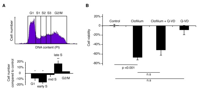
Figure 2. Inhibition of Erg activity results in a mainly apoptosis independent cell death in G1 and early S phase.
(A) mESCs were exposed to cisapride (10 µM) or vehicle for 6 h and DNA content was assayed using propidium iodide labeling by flow cytometry and quantified in respective cell cycle stages (one-way ANOVA, * p<0.05, ** p<0.01). (B) mESCs were treated with the Erg inhibitor, E4031 (10 µM), for 24 h with and without apoptosis inhibitor, Q-VD-OPh (20 µM) and viability was measured using an ATP detecting viability assay. Data presented as mean ± SEM (N=3), one-way ANOVA, Tukey post-hoc test.

