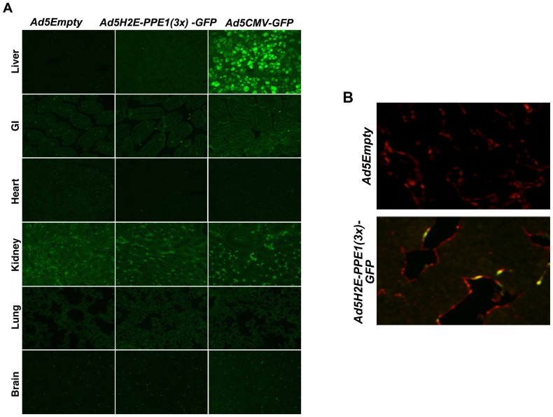
Figure 4. Intravenous administration of Ad5H2E-PPE1(3x)-GFP results in GFP expression selectively in tumor endothelium.
1×10¹⁰ PFU of Ad5Empty, Ad5H2E-PPE1(3x)-GFP or Ad5CMV-GFP were intravenously administered to MCA/129 fibrosarcoma-bearing sv129/BL6 mice. Five days post viral administration, normal tissues (A) and tumor tissue (B) were excised and GFP expression was visualized by standard fluorescence microscopy following staining with anti-GFP (green; A and B) and anti-Meca-32 (red; B) antibodies, as described in Materials and Methods. Shown are representative 200× images of 20 fields analyzed per sample. Note background autofluorescence in the kidney specimens.

