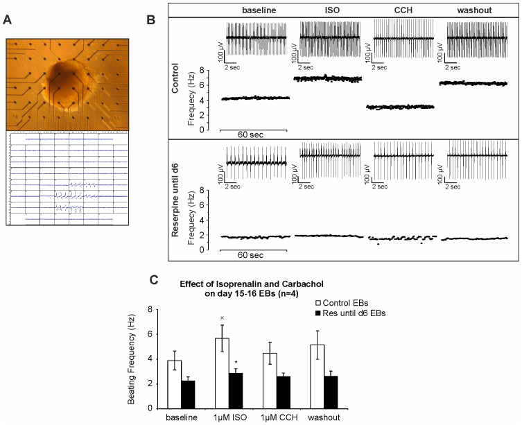
Figure 5. Comparison of β-adrenergic and muscarinic modulation of beating rate in control and reserpine-treated ES cell-derived cardiomyocytes.
(A) Spontaneously contracting EB plated on MEA. (B) Representative beating frequencies demonstrating effects of ß-adrenergic agonist isoprenaline (ISO, 1 µM) and muscarinic agonist carbachol (CCH, 1 µM) on cardiac clusters generated under control (top panel) and reserpine-treated (bottom panel) conditions. Original FP traces (10 sec) of each indicated condition are showcased (on top of each plot). (C) Statistical analysis of FP frequencies in MEA measurements (n = 4). (xp<0.05: significant difference between baseline and 1 µM ISO in ctrl EBs; *p<0.05: significant difference between ctrl and reserpine-treated EB under ISO (1 µM)).

