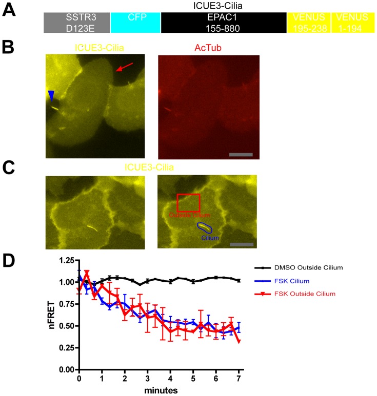
Figure 2. A FRET-based biosensor for local analysis of cAMP dynamics in individual cells.
A. Schematic representation of ICUE3-Cilia design. The biosensor consists of a ligand binding-defective SSTR3 to achieve ciliary localization fused to ICUE2 except with the C-terminal citrine present in ICUE2 replaced by a circularly permuted YFP variant to increase the overall fluorescence signal. B. Representative fluorescence micrograph of IMCD3 cells transfected with ICUE3-Cilia, showing YFP fluorescence (left panel, yellow) relative to AcTub immunoreactivity (right panel, red). ICUE3-Cilia localized both to the primary cilium (arrowhead) and was detectable in the extra-ciliary plasma membrane (arrow). Scale bar, 10 um. C. YFP image of a representative ICUE3-Cilia -transfected cell (left panel), with selected regions of plasma membrane outside of cilium (‘cell’, red outline) and including the primary cilium (‘cilia’, blue outline) indicated. nFRET determinations in each region were carried out as described in Experimental Procedures. Scale bar, 10 um. D. Time course of nFRET change in the indicated regions of IMCD3 cells expressing ICUE3-Cilia, with 5 uM forskolin or vehicle (DMSO) applied at t = 40 sec. The black line shows vehicle control indicating minimal photobleaching over the period of nFRET determination. The red line indicates extra-ciliary (‘outside cilium’) nFRET and blue line indicates ciliary (‘cilium’) nFRET calculated from the same image series (n = 3 experiments, ≥3 cells imaged per experiment, error bars indicate S.E.M. across experiments).

