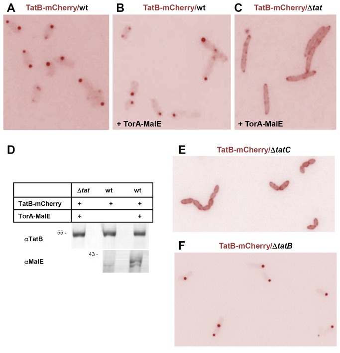
Figure 6. In the presence of TatA and TatC, TatB localizes almost exclusively to the cell poles.
(A) E. coli BL21(DE3) wild type cells (wt) expressing TatB-mCherry from a pBAD33 vector (pPR2) following 2 h of induction with 0.1% arabinose. The fluorescence signal accumulates predominantly in polar foci with almost no staining of the cell bodies. (B) Co-expression of TatB-mCherry from a pBAD33 vector (pPR2) as in (A) and of the Tat substrate TorA-MalE335 from a pET22 vector (pPJ1) at non-induced levels. The additionally expressed Tat substrate does not change the staining pattern of TatB-mCherry. (C) Co-expression of TatB-mCherry and TorA-MalE335 in a BL21(DE3) Δtat cells. Without TatA and TatC (Δtat), TatB-mCherry is found dispersed in the cell periphery rendering the shape of the cells now more discernible against the background. (D) Equal expression levels of TatB-mCherry in cells depicted in (A–C). Whole cell proteins were precipitated with trichloroacetic acid and equivalent amounts were each subjected to SDS-PAGE. Immunoblots were decorated with antibodies against TatB and MalE as indicated. (E) Expression of TatB-mCherry in a ΔtatC strain (B1lK0). With TatC missing but native levels of TatB in the cells, TatB-mCherry is distributed evenly in the membrane as observed in a ΔtatABC strain (C). (F) When TatB-mCherry is expressed in a ΔtatB strain (BΦD) with endogenous TatC present, a polar localization of TatB-mCherry is observed as in a TatABC wild-type strain (A, B).

