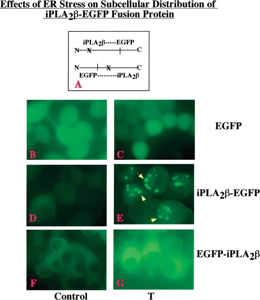
Figure 8.
Effects of ER stress on the subcellular distribution of an iPLA2β-EGFP fusion protein in INS-1 cells. (A) Schematic of two fusion protein constructs where X indicates the potential caspase-3 cleavage site in the iPLA2β protein. INS-1 cells were transfected with either EGFP vector alone (panels B and C) or constructs designed to attach the EGFP tag to the N-terminus (EGFP-iPLA2β, panels D and E) or the C-terminus (iPLA2β-EGFP, panels F and G) of iPLA2β. Cells were treated with vehicle only (Control, panels BD, and F) or with 1 µM thapsigargin (T, panels CE, and G) and examined by fluorescence microscopy 6 h later. The iPLA2β-EGFP-producing INS-1 cells treated with thapsigargin exhibit punctate perinuclear iPLA2β fluorescence, as indicated with yellow arrows (panel E).

