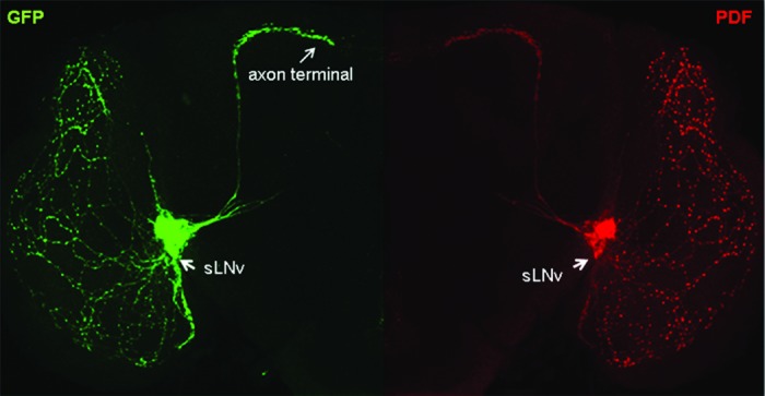
Figure 1.Drosophila ventrolateral neurons expressing GFP shown in green and the circadian neuropeptide pigment dispersing factor (PDF) shown in red. GFP and PDF Immunostaining label the circadian pacemaker cells, the small ventrolateral neurons (sLNv) including their dorsally projecting axons and terminal arbors in the central region of the adult fly brain. Axon growth and PDF expression in sLNv is epigenetically regulated by Tip60 HAT activity.
