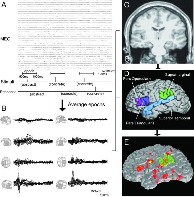
Fig. 1.
A, Schematic representation of language MEG processing. Stimuli are visually presented as “abstract” or “concrete” words (Stimuli line). Patient responses are also recorded (Response line). Using stimuli as a trigger, epochs from −500 ms to 1000 ms are averaged. B, Waveforms of the averaged MEG from 0 ms to 1000 ms. Waveforms of each MEG sensor group are superimposed. Language activation is seen around 250–550 ms in the frontal, temporal, and parietal sensors. MPRAGE MR imaging (C) provides the cortical surface of each patient by reconstruction processing. This procedure also gives anatomic parcellation of the cortex (D). Four ROIs per each hemisphere are used for calculating LI. E, Spatiotemporal source distribution of the language MEG is mapped on the cortical surface by using dSPM. The value of activation in ROIs is extracted from dSPM.

