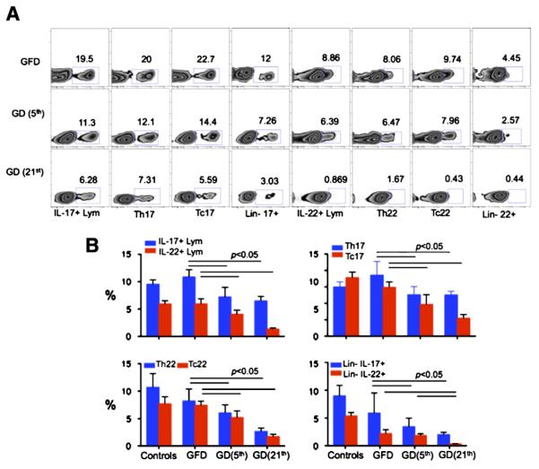
Figure 4.
Characterization of duodenal IL-17+ and IL-22+ cells. (A) Dietary gluten induced significant decrease of IL-17+/IL-22+ cells in duodenum of gluten-sensitive rhesus macaques. Differences in populations of IL-17+/22+ lymphocytes, IL-17+/22+CD4+ T helper (Th17+/22+) cells, IL-17+/22+CD8+ (Tc17+/22+) cells and IL-17+/22+ lineage-negative (Lin−) cells are shown concerning the GFD and GD periods. GD data are shown for days 5 and 21 following the introduction of GD. (B) Significant decreases of overall (group x ±STD) IL-17+/IL-22+ lymphocytes as well as Th, Tc and Lin− IL-17+/22+ cells are shown.

