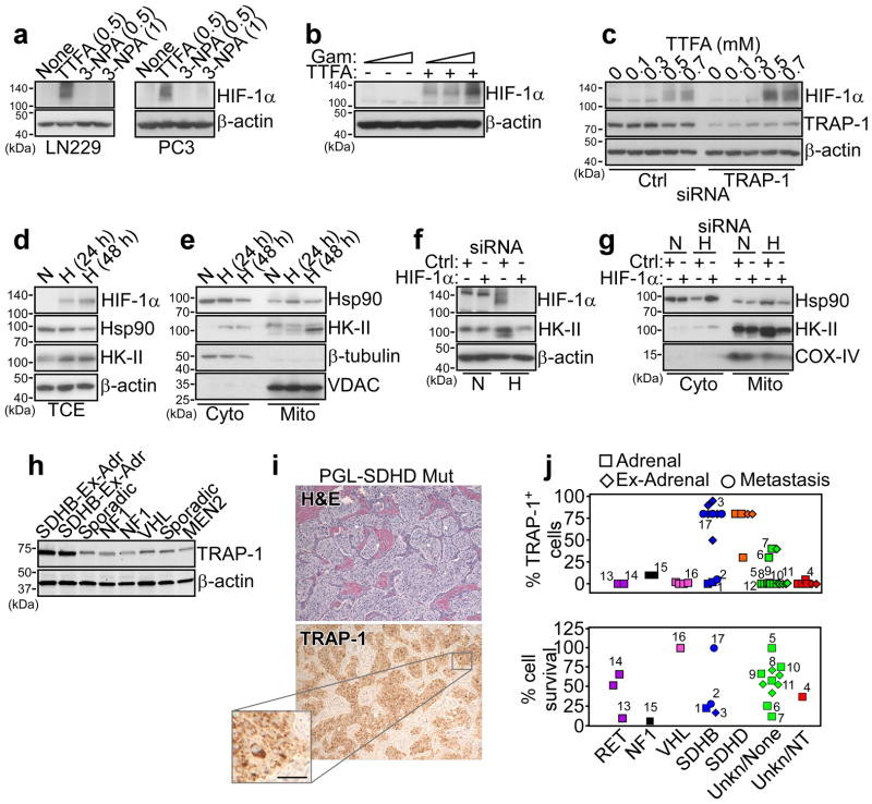
Figure 5. TRAP-1-SDHB complex regulates HIF-1α-directed tumorigenesis.
(a) The indicated tumor cell types (LN229 or PC3 cells) were treated with the various concentrations (mM) of the SDHB inhibitors, TTFA or 3-NPA and analyzed by Western blotting. (b) PC3 cells were treated with increasing concentrations of Gamitrinib (Gam, 0, 2.5, 5 μM) in the absence (−) or presence (+) of TTFA (0.3 mM) and analyzed by Western blotting. (c) LN229 cells were transfected with control siRNA (Ctrl) or TRAP-1-directed siRNA and analyzed by Western blotting in the presence of the indicated increasing concentrations of TTFA. (d, e) PC3 cells were maintained under conditions of hypoxia (H, 0.5% O2, 5% CO2, 94% N2 for 24 h) or normoxia (N), and analyzed by Western blotting in total cell extracts (TCE) (d), or fractionated cytosolic (Cyto) or mitochondrial (Mito) extracts (e). VDAC or β-tubulin was used as a mitochondrial or cytosolic marker, respectively. (f) PC3 cells were transfected with control siRNA (Ctrl) or HIF-1α-directed siRNA, maintained in normoxia (N) or hypoxia (H) conditions, and analyzed by Western blotting. (g) PC3 cells were transfected and treated as in (f), and isolated cytosolic (Cyto) or mitochondrial (Mito) fractions were analyzed by Western blotting. COX-IV was used as a mitochondrial marker. (h) Patient-derived tissue samples of PCC/PGL were analyzed by Western blotting. The mutational status of each tumor is indicated. Ex-Adr, extra-adrenal localization. (i) A tissue sample of extra-adrenal PGL with SDHD mutation, showing a typical nest-like (“Zellballen”) growth pattern was stained with hematoxylin/eosin (H&E, top) or TRAP-1 (bottom), by immunohistochemistry. Scale bar, 50 μm. (j) Quantification of immunohistochemical expression of TRAP-1 in PCC/PGL cases with the indicated mutational status (top). Cells from the various tumor samples were maintained in culture and analyzed for killing by Gamitrinib (10 μM for two weeks) (bottom) measured by counts of tyrosine hydroxylase-positive cells counted in an area defined by a randomly placed 22×22 mm square coverslips in 35 mm round culture dishes. Each point represents a single tumor. Paired samples of the same tumor were available in 12 instances and are indicated by matching numbers. Data are from a representative experiment.

