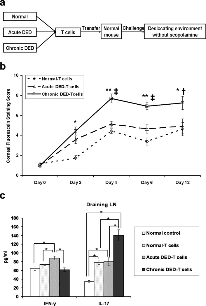
Figure 2.
T cells from chronic dry eye disease (DED) mice induce the most severe disease in naïve recipients. (a) Schematic study design of adoptive transfer experiment. Total T cells were isolated from normal, acute and chronic DED (n = 3–5 mice/group). Each naïve recipient was then injected i.v. with 1 × 10⁶ cells. Recipients were challenged in the controlled-environment chamber for 22 d without use of scopolamine. (b) Disease severity comparison among the recipients transferred with the different T cells. The mean corneal fluorescein staining score ± SEM for each group (n = 16 eyes) is shown. *, p < 0.05; **, p < 0.01 compared to normal-T cell recipients. †, p < 0.05; ‡, p < 0.01 compared to acute DED-T cell recipients. (c) T cell response at the recipient ocular surface. Conjunctival tissue was collected from the recipients at day 6, and relative mRNA expression of IFN-γ and IL-17 transcripts (n = 6 eyes per group) was quantified. Data shown are mean ± SEM. *, p < 0.05; **, p < 0.01. (d) T cell response in the recipient draining lymph nodes (LNs). Draining LNs were harvested at day 6, and analyzed for IFN-γ and IL-17 levels by ELISA (n = 3 mice per group). Data shown are mean ± SEM. *, p < 0.05. Groups are labeled as follows: Normal T-cells, recipients of T cells from normal mice; Acute DED-T cells, recipients of T cells from acute DED mice; Chronic DED-T cells, recipients of T cells from chronic DED mice.

