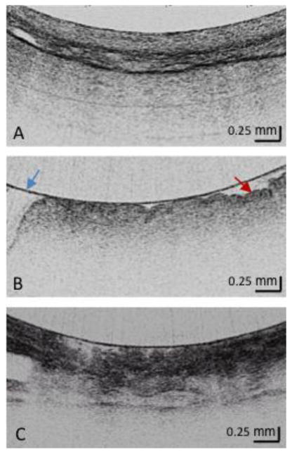
Figure 1. Squamous, Gastric Cardia and Barrett's Metaplasia via OFDI.

- Squamous Mucosa Layered structure No glands in the epithelial layer Squamous epithelium is homogeneous
- Gastric Cardia Mucosa (at least 2) Vertical pit architecture Highly reflective surface (red arrow) Relatively poor image penetration Broad regular foveolar region Rugae Blue arrow is the balloon wall
- Barrett's Metaplasia (at least 2) Loss of layered or vertical pit and crypt architecture Irregular mucosal surface Heterogeneous scattering
