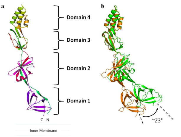
Figure 2.
Crystal structure of the CusB membrane fusion protein. (a) The structure can be divided into four distinct domains. Domain 1 is formed by the N and C-termini and is located above the inner membrane. The loops between domains 2 and 3 appear to form an effective hinge to allow the molecule to shift from an open conformation to a more compact structure. Domain 4 is folded into an anti-parallel, three-helix bundle, which is thought to be located near the outer membrane. (b) This figure depicts a comparison of the two conformations of CusB observed in the crystal. Superimposition of domains 3 + 4 of the two crystal structures of CusB displays an overall shift of the -strands of domain 1 of CusB by ~23°.

