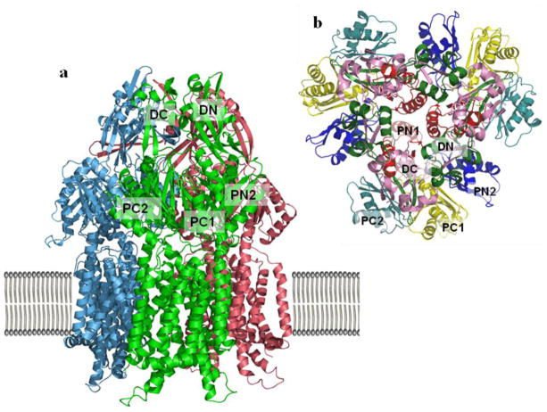
Figure 3.
Crystal structures of the CusA efflux pump. (a) Ribbon diagram of the CusA homotrimer viewed in the membrane plane. Each molecule is labeled with a different color: molecule 1 is green, molecule 2 is pink, and molecule 3 is cyan. Each subunit of CusA consists of 12 transmembrane helices (TM1-TM12) and a large periplasmic domain formed by two periplasmic loops between TM1 and TM2, and TM7 and TM8, respectively. (B) Ribbon diagram of the CusA homotrimer subdomains as viewed from the top. The six subdomains are PN1 (red), PN2 (blue), PC1 (yellow), PC2 (teal), DN (green) and DC (pink).

