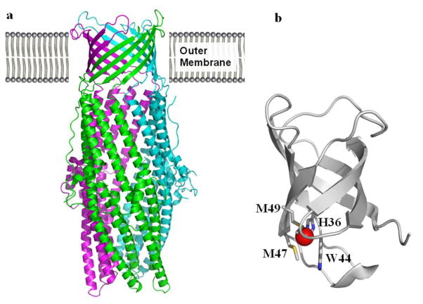
Figure 8.
Crystal structures of CusC and CusF. (a) Ribbon diagram of the CusC homotrimer (pdb code: 3PIK) (Kulathila et al. 2011) viewed in the membrane plane. Each molecule is labeled with a different color (green, pink, and cyan). (b) Ribbon diagram of Cu(I)-bound CusF (pdb code: 2VB2) (Xue et al. 2008). H36, M47, M49 and W44 forming a metal binding site are shown in stick form. The bound Cu(I) is in red.

