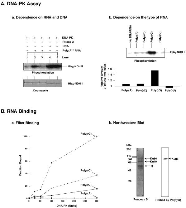
Figure 6.
RNA-dependent phosphorylation of NDH II by DNA-PK. (A) (Aa) DNA-PK phosphorylated NDH II in the presence of native RNA. Purified His6-tagged NDH II (≈1 µg each) (23) was used as protein substrate for the DNA-PK assay. Poly(A)+-RNA was from mouse spleen (Clontech) and added to ≈0.1 mg/ml. DNA was from calf thymus and used at 0.05 mg/ml. RNase A treatment was as described in Figure 5. (Ab) Phosphorylation of NDH II in the presence of synthetic RNAs. DNA-PK was incubated with His6-tagged NDH II (≈1 µg each) in the presence of poly(rA), poly(rC), poly(rG) and poly(rU). Phosphorylation levels relative to the background (i.e. in the absence of nucleic acids) are also presented. The signals were quantified with a PhosphorImager. (B) RNA binding assay of DNA-PK. (Ba) Filter binding of DNA-PK with the indicated 32P-labeled ribopolymers poly(rA), poly(rC), poly(rG) and poly(rU). (Bb) Northwestern blot of the Ku heterodimer. Ku86/70 was obtained from HeLa nuclear extract by immunoprecipitation as described in Figure 1. After separation by SDS–PAGE the proteins were transferred to a Hybond-C nitrocellulose membrane (Amersham) and probed by the same RNAs as used for filter binding. Only the Ponceau S protein staining and the membrane probed with poly(rG) are shown. Ig, immunoglobulin.

