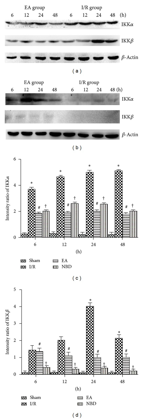
Figure 10.

Western blot analysis of the IKKα and IKKβ protein expression in the sham group, I/R group, EA group, and NBD group in ischemic brain tissue (with the same samples from Figure 7). (a)-(b) Representative Western blot images showing bands of IKKα and IKKβ proteins at 6 h, 12 h, 24 h, and 48 h. Comparison of the mean intensity ratio of immunoblotting in these groups at each timepoint. (a), (b), (c) The IKKα protein in I/R group was highly expressed, *P < 0.05 versus EA and NBD group. The expression in EA group was lower, # P < 0.05 versus NBD group. (a), (b), (d) The IKKβ protein was highly expressed at 24 h and 48 h, *P < 0.05 versus EA and NBD group. The IKKβ protein scarcely expressed in NBD group, † P < 0.05 versus EA group.
