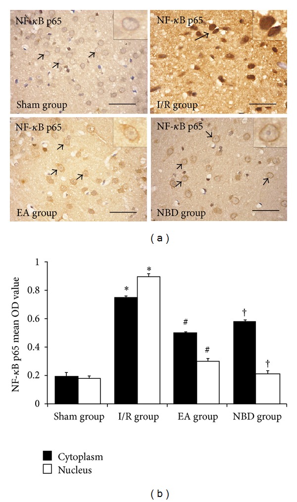
Figure 4.

Immunohistochemical analysis of the nuclear translocation of NF-κB p65 in the sham, I/R group, EA group, and NBD group (5 rats in each group, total 20 rats). In the temporal neocortex of sham group, the immunoreactive staining occurred less in the cytoplasm and nucleus. In the I/R group, strong immunoreactive staining occurred in the cytoplasm and nucleus, especially in the nucleus, *P < 0.05. In the EA group, immunoreactive staining was predominantly detected in the cytoplasm rather than in the nucleus, # P < 0.05. In the NBD group, immunoreactive staining was also maintained in the cytoplasm, † P < 0.05. Comparison of the mean OD value showed that NF-κB p65 was mainly expressed in the nucleus after focal ischemia/reperfusion, and expression of NF-κB p65 protein in the nucleus in the EA group and NBD group was significantly reduced, # P < 0.05, † P < 0.05.
