Figure 3.

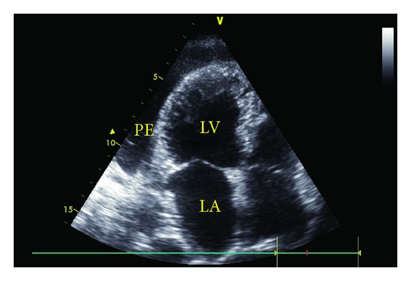
Apical four-chamber view with transthoracic echocardiography (PE: pericardial effusion, LV: left ventricle, and LA: left atrium).

Apical four-chamber view with transthoracic echocardiography (PE: pericardial effusion, LV: left ventricle, and LA: left atrium).