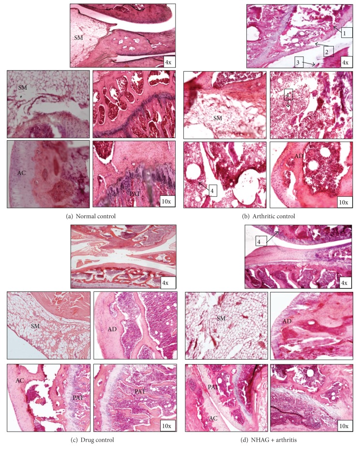
Figure 5.
Histology of knee joints from arthritic treated, nontreated, and normal animals (hematoxylin and eosin staining). AC: articular cartilage; AD: articular disc; SM: synovial membrane; PAT: periarticular tissue. Normal control: note lack of lymphocytes infiltration of synovium and intact articular bone. Arthritic control: prominent lymphocytic infiltration of synovium with invasion of periarticular bone and vacuolization (arrow 1 and 4), collapse of articular surface, and articular bone destruction (arrow 2 and 3). Drug control: note mild infiltration of lymphocytes in synovium, however; the damage in articular bone is quite negligible. NHAG + arthritis: note mild to moderate infiltration of lymphocytes in synovium with slightly damaged articular bone (arrow 4).

