Fig. 4.

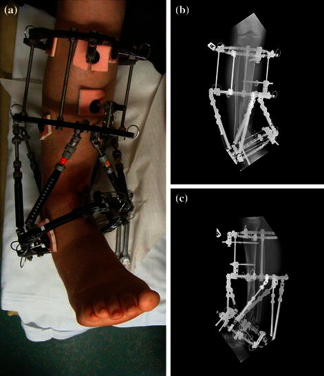
Waiting for the soft tissue envelope to heal. The intentional varus and recurvatum are seen with the TSF in situ. a Clinical picture. b, c AP & Lateral view of the intentional deformity

Waiting for the soft tissue envelope to heal. The intentional varus and recurvatum are seen with the TSF in situ. a Clinical picture. b, c AP & Lateral view of the intentional deformity