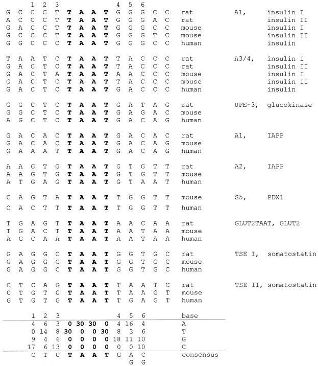
Figure 2.
Tabulation of native PDX1 binding sites. Alignment of 30 sequences of experimentally defined potential PDX1 binding sites from human, mouse and rat genes relative to the TAAT motif (shown in bold). Designations of the PDX1 binding sites [according to nomenclature from (39,41,42,44–46)] are indicated on the right together with the corresponding genes. TAAT flanking positions are numbered. Below the sequences, a nucleotide frequency matrix and the derived consensus are shown.

