
Fig. 1.
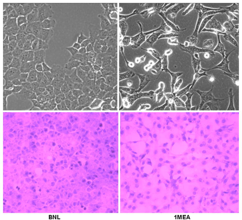
BNL and 1MEA cells are morphologically different. BNL cells display typical epithelial morphology whereas 1MEA cells show mesenchymal characteristics. Image magnification was ×40 objective for unstained cells and ×20 objective for H&E stained cells. Left BNL cells, right 1MEA cells