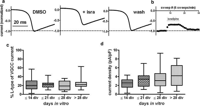
Fig. 9.
Levels of LTCC-mediated calcium currents in primary hippocampal neurons. a LTCC-mediated current components in total voltage-gated calcium currents were determined by applying ramp depolarizations (0.5 mV/ms) from −80 mV (=holding potential) to +50 mV and measurement of calcium current reduction upon a 90-s administration of 3 μM isradipine. The three traces depict the peak currents evoked under control conditions (DMSO), 3 μM isradipine and after washout of the dihydropyridine. b The reversible reduction was monitored by reading the peak of currents that were elicited every 10 s (e.g., sweeps 8–16 in the experiment shown). c Percentage of isradipine inhibited current with respect to total voltage-activated currents calculated from measurements as shown in a, b. Neurons were grouped according to the age of the cultures, as indicated on the x-axes. Neurons that had been kept in culture for at least 10 days but not longer than 2 weeks were allocated to the ≤14 days in vitro (DIV) group (n = 16), neurons that had been maintained in culture for more than 4 weeks and maximally up to 5 weeks were allocated to the >28 DIV group (n = 19). n for the ≤21 DIV and ≤28 DIV was 17 and 15, respectively. Considerably variation of LTCC current density exists in all age groups, yet statistically groups do not significantly differ from each other. d Same data as in c. LTCC current density (pA/pF) was determined by relating of the dihydropyridine-sensitive current component to cell capacitance as a measure of cell surface. To highlight the intrinsic variation, data in c and d are shown as box-plots with min to max whiskers

