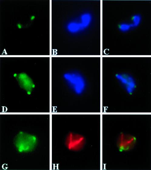
Figure 6.
Spo21-GFP localizes to the spindle pole. Strain AN230 (spo21::GFP/spo21::GFP) carrying pRS316-SPO21::GFP2 was transferred to 2% KOAc and analyzed by fluorescence microscopy as described in MATERIALS AND METHODS. (A) Spo21-GFP in a meiosis I cell. (B) DAPI staining of the same cell in A. (C) Merged image of A and B showing localization of Spo21-GFP to the periphery of the nucleus. (D) Spo21-GFP foci in a meiosis II cell. (E) DAPI staining of the same cell in D. (F) Merged image of D and E showing Spo21-GFP near the leading edge of the segregating chromatin. (G) A meiosis II cell showing four Spo21-GFP foci. (H) Antitubulin staining of the same cell in G. (I) Merged image of G and H, demonstrating the Spo21-GFP foci are at the spindle poles.

