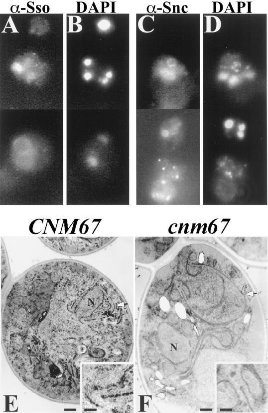
Figure 9.
Prospore membranes form in abnormal locations in a cnm67 mutant. Strain AN161 (cnm67Δ/cnm67Δ) was transferred to 2% KOAc and prepared for immunofluorescence and transmission EM as described in MATERIALS AND METHODS. (A) Anti-Ssop staining. (B) DAPI staining of cells in A. (C) Anti-Sncp staining. (D) DAPI staining of cells in C. (A–D) Composite images. (E) Transmission EM of a wild-type cell prepared by permanganate staining shows a prospore membrane engulfing a nuclear lobe (arrowhead). (F) A cnm67 mutant prepared similarly to the cell in E displays a prospore membrane (arrowhead) unassociated with nucleus (N). Insets: Higher magnification showing the characteristic knob on the end of the prospore membranes in E and F. Scale bars: large panels, 500 nm; insets, 150 nm.

