Figure 9.

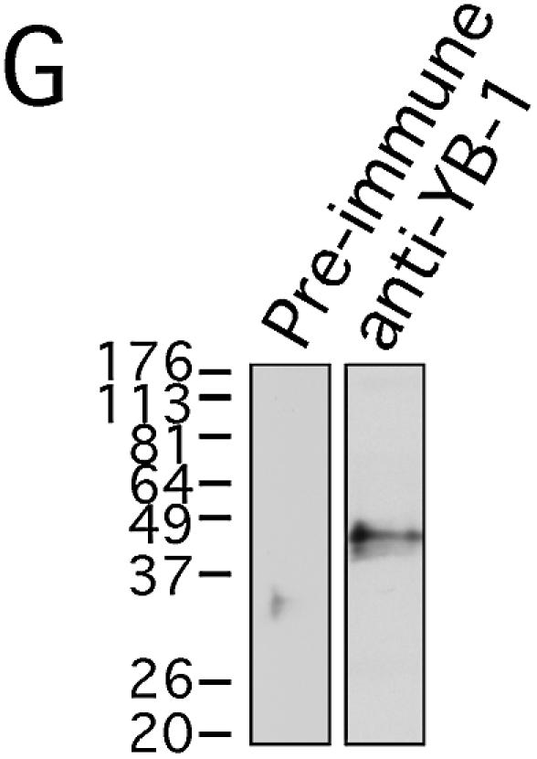
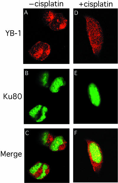
Co-localization of YB-1 and Ku80 by immunofluorescence. Human 293 embryonic kidney cells were untreated (A–C) or incubated with 7.5 µg/ml of cisplatin for 2 h (D–F), then fixed and subsequently incubated simultaneously with rabbit anti-YB-1 and mouse anti-Ku80 as described in Materials and Methods. Anti-rabbit rhodamine-labeled and anti-mouse FITC-conjugated secondary antibodies were used to visualize YB-1 and Ku80 by confocal microscopy at 568 and 488 nm, respectively. Images depict representative cells from each of the non-treated and cisplatin-treated cultures. In the merged images (C and F) a yellow color appears where YB-1 (red) and Ku80 (green) fluorescence signals coincide. In (G), western analysis of 293 total cell lysate with anti-YB-1 and pre-immune sera.

