Figure 2.
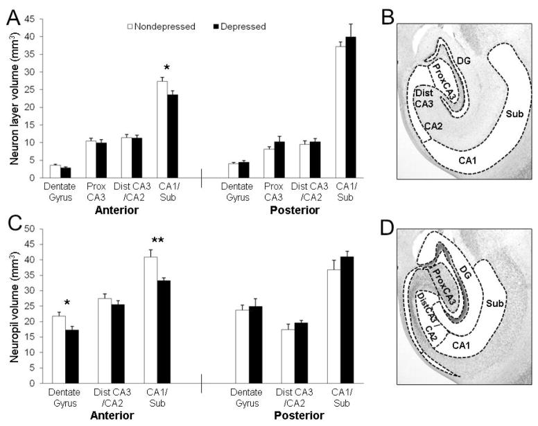
CA1 and DG neuropil and principle cell layers were smaller in the anterior hippocampus of depressed monkeys. (A) No main effect of depression was found, but there was an interaction by subregion (F(3,42) = 4.32, p < 0.01) for principle cell layer volumes in the anterior hippocampus, with reduced volume of the pyramidal cell layer in the CA1/subiculum (p < 0.05) of depressed monkeys. No effects were observed in the posterior hippocampus. (B) The subregion delineations for principle cell layers are depicted. (C) Neuropil volumes were smaller in depressed monkeys (F(1,14) = 8.41, p < 0.05). The depression × subregion interaction was suggestive but did not reach significance (F(3,42) = 3.22, p < 0.06). Smaller neuropil extent was observed in the anterior CA1/subiculum (p < 0.01) and DG (p < 0.05). No effects were found in the posterior hippocampus. (D) The subregion delineations for neuropil layers are depicted. *p < 0.05; **p < 0.01.

