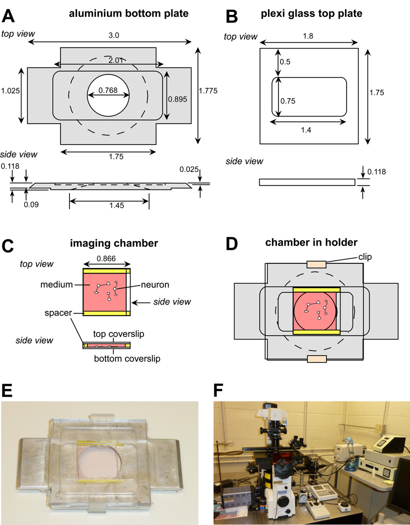
Figure 1. Imaging chamber used for live cell imaging of neuronal growth cones.
(A) Schematic drawing of bottom aluminium plate of custom-made holder used for the imaging chamber described in this chapter. Dimensions are in inches. (B) Plexiglass top plate of custom-made holder. (C) Imaging chamber made by two coverslips separated by two plastic spacers. The bottom coverslip contains the cultured neurons. Left and right side are open for medium exchange. (D) Imaging chamber mounted between bottom and top part held together by plastic clips. (E) Picture of an assembled chamber within the holder. (F) Live cell imaging workstation based on an Eclipse TE2000E2 (Nikon) inverted microscope as described in this article.

