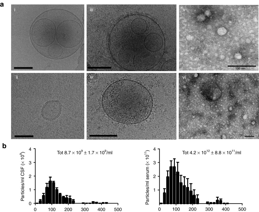
Figure 3.
Extracellular particle analysis using EM, cryoTEM and Nanosight. Cerebrospinal fluid (CSF), plasma and serum samples were collected upon or before the opening of the dura mater, respectively, processed as described in methods and stored at −80 °C until further processing. (a) CryoTEM of CSF EVs (i, ii), plasma EVs (iii, iv), and regular EM of CSF EVs (v, vi). Scale bars = 100 nm. (b) CSF (n = 3) and serum samples (n = 3) were diluted 1:50 and 1:3,000 respectively in PBS, and size and concentration of particles were assessed using nanoparticle tracking analysis (NanoSight; ±SD).

