Fig. 3.
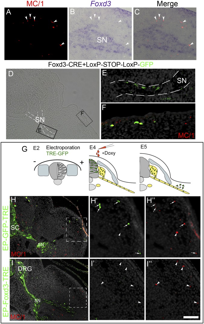
Sustained Foxd3 activity inhibits melanocyte differentiation of bipotent SCP–melanocyte progenitors. (A–C) Transverse section of an E5 limb showing nonoverlapping Foxd3 and MC/1 signals in SCPs along a spinal nerve and melanocytes (arrowheads), respectively. (D–F) MC/1+ melanocytes develop from Foxd3+ SCPs. Staining for MC/1 of a section through the hindlimb-level of an E5.5 embryo whose dorsal NT was focally electroporated at E2 with enhancer #168-cre and pCAGG::LoxP-STOP-LoxP-GFP. (E and F) Magnifications of boxed areas in D showing enhancer-positive cells along the nerve (E) and in the dermis/epidermis (F) where they coexpress MC/1. Nerve is highlighted with dashed white lines. (G–I) Inducible expression of Foxd3 in SCP along spinal nerves. (G) Experimental scheme: electroporation of pCAGGS-rtTA2s-M2 along with either pBI-TRE-GFP or pBI-TRE-Foxd3 into hemi-NT at hindlimb levels of E2 embryos. Doxycyclin was injected 2 d later, and the localization and fate of transfected cells was assessed at E5. In control embryos, MC/1+ cells in the limb are GFP+ (arrows, H–H’’’), whereas MC/1+ melanocytes in treated embryos are Foxd3/GFP-negative (arrowheads, I–I’’’). Doxy, doxycycline; SC, spinal cord. (Scale bar, 50 μm in A–C; 70 μm in D; 30 μm in E and F; 80 μm in H and I; 40 μm in H’–H’’’ and I’–I’’’.)

