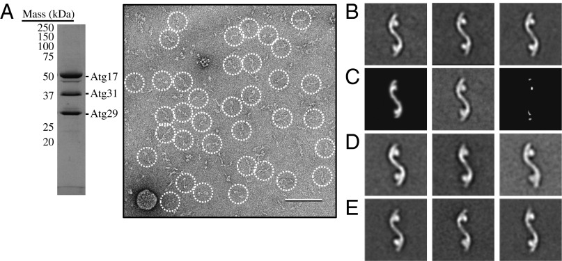
Fig. 5.
The Atg17-Atg31-Atg29 complex adopts an elongated S-shaped structure. (A) Coomassie blue–stained SDS/PAGE analysis of gel filtration-purified recombinant Atg17-Atg31-Atg29 (Left). A raw image taken from a negatively stained recombinant Atg17-Atg31-Atg29 specimen (Right). Particles are circled. (Scale bar, 100 nm.) (B) Representative class averages obtained from reference-free classification of 6,788 negatively stained Atg17-Atg31-Atg29 particles into 50 classes. Each of the three classes contains between 110 and 220 particles. The side length of each panel is 52 nm. (C) Difference mapping between the high-resolution crystal structure projection (Left) and the negative-stained EM average of the full-length components from S. cerevisiae (Center). The difference image (Right) indicates that the Atg29 C terminus is located in the globular domains. (D) Representative class averages obtained from classification of 4,671 negatively stained Atg29[3SD]-containing particles into 50 classes. Each of the three classes contains between 100 and 190 particles. (E) Representative class averages obtained from classification of 6,588 negatively stained Atg29[20STA/3SD]-containing particles into 50 classes. Each of the three classes contains between 150 and 170 particles.

