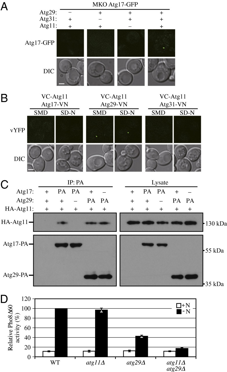
Fig. 6.
Atg11 is important for PAS recruitment of the Atg17-Atg31-Atg29 complex. (A) An empty vector or a plasmid expressing Atg29-PA was coexpressed with empty vector or a plasmid encoding HA-Atg11 under the control of the CUP1 promoter (pCuHA-Atg11) in MKO ATG17-GFP (HCY107) or MKO ATG17-GFP atg31∆ (HCY108) cells as indicated. Cells were cultured in SMD to early log phase. (B) VC-ATG11 ATG17-VN (KDM1551), VC-ATG11 ATG29-VN (KDM1552), and VC-ATG11 ATG31-VN (KDM1553) cells were cultured in YPD and shifted to SD-N for 2 h. All of the cell samples in A and B were observed by fluorescence microscopy. The images are representative pictures from single Z-sections. DIC, differential interference contrast. (Scale bar, 2 μm.) (C) The plasmid pCuHA-Atg11(416) was transformed into WT (SEY6210), ATG17-PA (KDM1234), ATG17-PA atg29Δ (KDM1235), ATG29-PA (HCY129), or ATG29-PA atg17Δ (HCY125) cells. Cells were cultured in SMD, and cell lysates were prepared and incubated with IgG-Sepharose for affinity isolation as described in Materials and Methods. The eluted proteins were separated by SDS/PAGE and detected with monoclonal antibody that recognizes HA or PA. (D) Pho8Δ60 WT (TN124), atg11Δ (KDM1406), atg29Δ (YIY36), or atg11Δ atg29Δ (KDM1407) cells were cultured in YPD (+N) and shifted to SD-N for 4 h. The Pho8Δ60 assay was performed. Error bars correspond to the SD and were obtained from three independent repeats.

