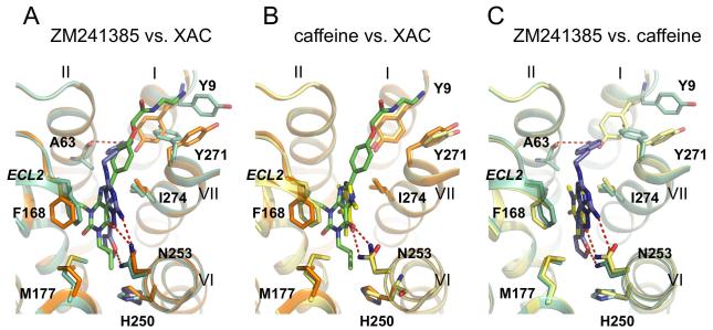
Figure 7. Comparison of specific interactions in the ligand binding site between the A2A-StaR2-ZM241385/XAC/caffeine co-structures.
a) Superposition of A2A-StaR2 (pale green) bound to ZM241385 (blue) with A2A-StaR2 (orange) bound to XAC (green) b) Superposition of A2A-StaR2 (orange) bound to XAC (green) with A2A-StaR2 (yellow) bound to caffeine (bright yellow) c) Superposition of A2A-StaR2 (pale green) bound to ZM241385 (blue) with A2A-StaR2 (yellow) bound to caffeine (bright yellow). A2A-StaR2 is shown in helical representation with specific side chains making important ligand receptor interactions marked. Specific hydrogen bonds are marked as dashed red lines. Ligands are represented as stick models (coloured according to the ligand) with oxygen atoms red and nitrogen atoms blue.

