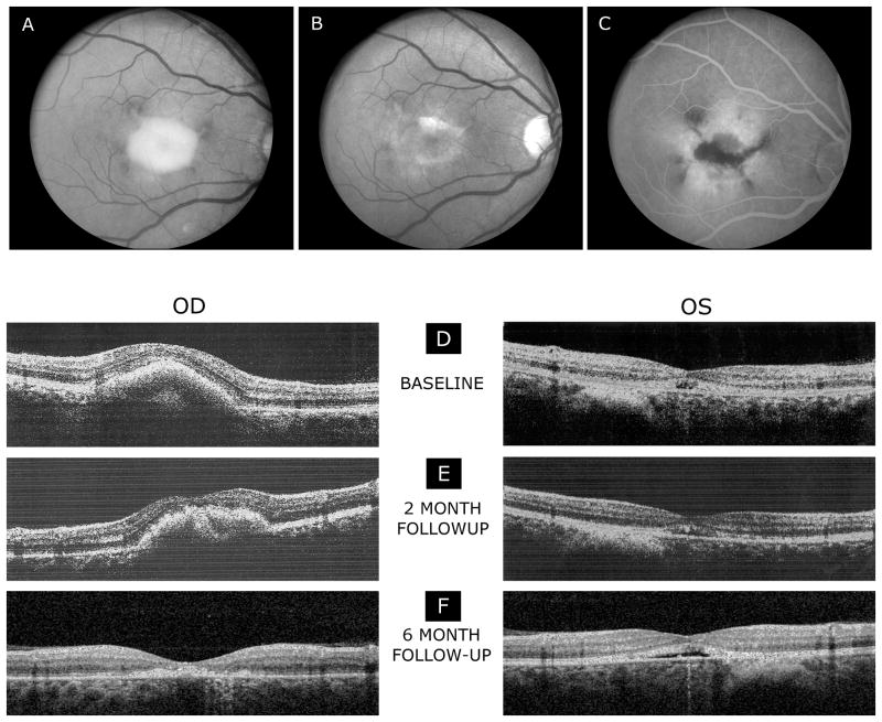
Fig.1.
Fundus photographs of the right eye (A), demonstrating an elevated vitelliform macular lesion with hyperpigmented changes at the margins. After six months of treatment (B), the vitelliform macular lesion in the right eye was less apparent when compared to the baseline. The right eye showed a small residual yellowish lesion in the macula with pigment on the surface of the lesion. Fluorescein angiogram (late frames) of the right eye (C) shows window defects of hyperfluorescence in the macula with hypofluorescent loci centrally. Spectral-domain OCT scans (E-F) demonstrate a reduction of the vitelliform macular lesion and retinal thickness while on treatment with topical brinzolamide 1% following two (E) and six months (F) of treatment. The left eye shows a shallow RPE elevation most evident at 6 months visit.

