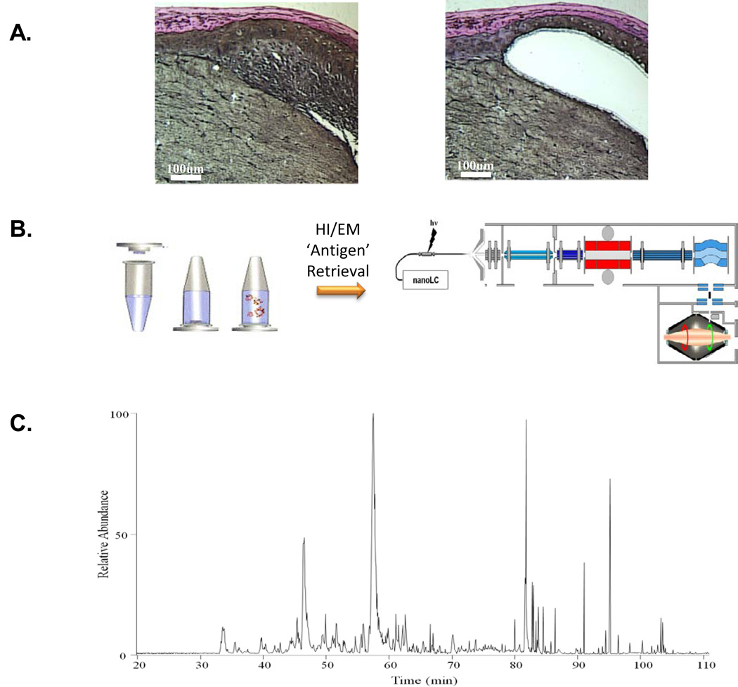
Figure 2. Mass spectrometry-based proteomic analysis of melanoma and skin organ cultures.
A. Hematoxylin & eosin staining of formalin-fixed, paraffin-embedded (FFPE) skin organ culture (SOC) sections injected with approximately 10,000 VGP melanoma cells (WM983-A) before (left panel) and after (right panel) laser microdissection. C. Basepeak chromatogram of approximately 5,000 VGP melanoma cells obtained by laser microdissection from the FFPE SOC tissue sections.

